Femoral Acetabular Impingement – FAI
HIP ARTHROSCOPY: FEMORAL ACETABULAR IMPINGEMENT – FAI
Femoral Acetabular Impingement – FAI – is a cause of early osteoarthritis of the hip in young and active people 20-40 years old. It is a result of too much friction in the hip joint. Basically, the ball (femoral head) and socket (acetabulum) rub abnormally creating damage to the hip joint. This causes pain and eventual loss of motion of the hip. The damage can occur to the articular cartilage (smooth white surface of the ball or socket) or the labral cartilage (soft tissue bumper of the socket).
FAI typically occurs during flexion and internal rotation. You will feel groin pain with hip rotation, in the sitting position, and/or during/after sports such as hockey, football, soccer, rugby, martial arts, tennis, golf, and/or baseball. You may be aware of decreased hip range of motion long before you feel pain. Pain can be minimal with straight and level walking. You may feel catching/clicking with movement of your hip – i.e. mechanical symptoms. While the cause is not well understood, patients with FAI often complain of low back pain. This pain is often localized to the SI joint (sacroiliac joint on back of pelvis), the buttock, the groin, and/or the greater trochanter (side of hip). FAI pain typically does not go beyond the level of the knee.
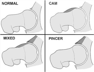
The two basic mechanisms of FAI are cam impingement (most common in young athletic males) and pincer impingement (most common in middle-aged women). This classification is based on the type of anatomical variation contributing to the impingement process. Cam impingement is the result of abnormal shape of the femoral head-neck junction; while pincer impingement is the result of an abnormal shape of the hip socket (acetabulum). In this situation the socket “pinches” the neck during hip movement. Cam lesions on the femoral head lead to shear forces of the non-spherical portion of the femoral head against the acetabulum resulting in a characteristic pattern of anterosuperior cartilage loss over the femoral head and corresponding dome, as well as labral tears. Labral tears associated with cam impingement are more commonly affect the transition zone cartilage and leaving the labral tissue in fairly good condition. Pincer lesions result in degeneration, ossification and tears of the anterosuperior labrum, as well as the characteristic posteroinferior “contre-coup” pattern of cartilage loss over the femoral head and corresponding acetabulum. In this setting, the acetabular labrum fails first which leads to degeneration and eventual ossification, which worsens the over coverage. Overall, the pincer type lesion has limited chondral damage compared to the deep chondral inury associated with cam impingement. FAI is mixed (Cam + Pincer) 86% of the time.
Femoral Acetabular Impingement
- Acetabulum (Excessive Coverage) = Pincer Impingement
- General
- Coxa Profunda
- Protrusio Acetabuli
- Focal
- Anterior (Acetabular Retroversion)
- Posterior (Prominent Posterior Wall)
- General
- Femur (Non-Spherical Head) = Cam Impingement
- Osseous Bump
- Lateral (Pistol Grip Deformity)
- Anterosuperior
- Femoral Retroversion, Coxa Vara
- Osseous Bump
Most patients can be diagnosed with a history, physical exam, and plain x-ray films. The physical exam will generally confirm the patient’s history and eliminate other causes of hip pain. A thorough evaluation of the lumbosacral spine (sacroilitits, lumbar spine degenerative disc disease), abdomen (hernia, abdominal aortic aneurysm), knee (arthritis or malaignment), and neurovascular examination (periperharl vascular disease) of the affected extremity will be performed to exclude peripheral sources of referred pain. The plain x-ray films are used to determine the shape of the ball and socket as well as assess the amount of joint space in the hip. Less joint space is generally associated with more arthritis. Further testing, such as a MRI and/or a diagnostic hip injection, may be indicated.
The treatment of FAI is based on how much arthritis is present in the hip joint. If there is severe arthritis, with near bone-on-bone disease, then a joint replacement would be indicated. However, a typical patient with FAIS has early arthritis and bone spurs (osteophytes) that are abnormally rubbing against each other causing labral tearing and injury to the cartilage. Surgery is most commonly performed to remove the bone spurs, sometimes referred to as the “hump” by contouring the bone on the neck (procedure called a osteoplasty). Surgery also includes removing hyaline cartilage that is diseased (chondroplasty) and repair or removal of the injured labrum
Originally the surgery was described as a surgical dislocation of the hip. This procedure is commonly performed, however, with the better technology Dr. Langer can perform the surgery using an arthroscope (hip arthroscopy). The excess bone is then removed under direct visualization and the labrum is repaired if possible. The arthroscopic approach involves less post-operative morbidity and allows patients, including professional athletes, to return to high-functioning lifestyles.
Recovery time is dependent on the patient – typically 3-6 months. Please see our videos on FAI, Hip Surgery Protocol, and Hip Arthroscopy Physical Therapy for more details.
To learn more about FAI and various conditions that can affect the hip please contact Dr. Langer for a consultation

